
Figure 3.
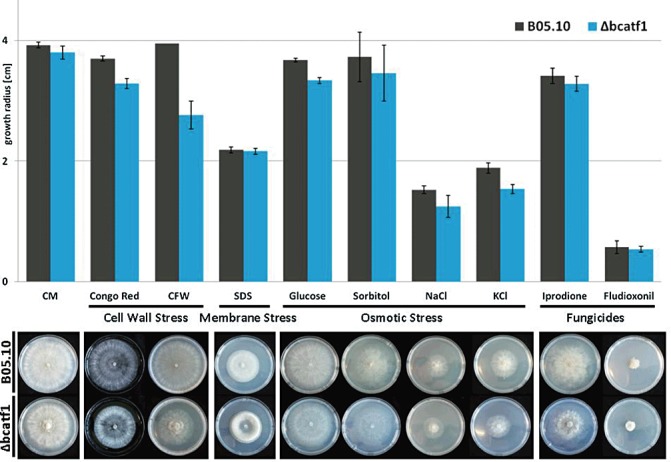
Growth in the presence of various stressors. Growth of Δbcatf1 and the wild‐type (WT) was tested on stress‐inducing media (3 days post‐inoculation). Complete medium was supplemented with Congo red (2 mg/mL), calcofluor white (CFW) (2 mg/mL), sodium dodecylsulphate (SDS) (0.02%), glucose (1 m), sorbitol (1 m), NaCl (1 m), KCl (1 m), iprodione (0.1 µg/mL) and fludioxonil (0.01 µg/mL). (A) Bar chart (growth radius in centimetres, mean value of three biological replicates, standard error is indicated). (B) Representative plates.