Figure 4.

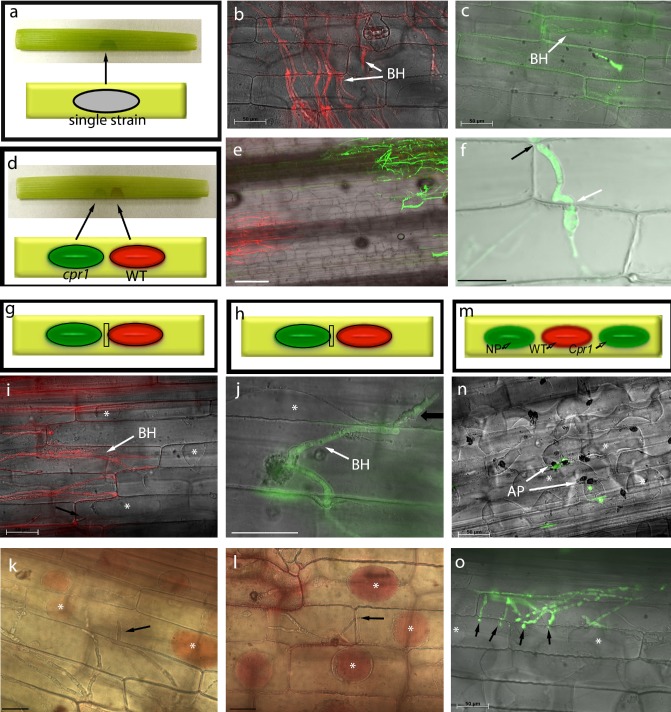
Diagrams of leaf sheath inoculations (a) and co‐inoculations (d, g, h, m). Colonization by WT‐mRFP (b) and cpr1‐Zsgreen (c), at 72 h post‐inoculation (hpi), on maize leaf sheaths. (e) cpr1‐Zsgreen colonization in co‐inoculations with WT‐mRFP. (f) Confocal image showing cpr1‐Zsgreen crossing intact plant cell walls (arrows) in co‐inoculations. Viability of newly invaded plant cells (arrows) and of cells surrounding wild‐type (WT) (i, k) and cpr1 (j, l) colonies in co‐inoculations, demonstrated by plasmolysis and neutral red staining (asterisks), at 60 hpi. (m–o) Triple inoculations with WT‐mRFP, cpr1‐Zsgreen and nonpathogen (NP) CgSl1‐GFP. (n) Nonpathogen failed to penetrate maize tissue, and cells underneath appressoria continued to plasmolyse (asterisks). (o) cpr1‐Zsgreen invading adjacent cells (arrows) that are still alive (asterisks). Scale bars (except f), 50 μm; (f) 20 μm. AP, appressoria; BH, biotrophic primary hyphae.
