Figure 2.

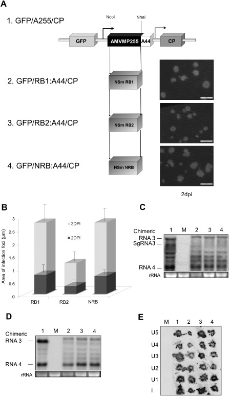
Analysis of the accumulation, cell‐to‐cell and systemic transport of the Alfalfa mosaic virus (AMV) chimeric RNAs carrying the movement protein (MP) of Tomato spotted wilt virus (TSWV) isolates. (A) Infection foci observed in P12 plants inoculated with RNA 3 transcripts from pGFP/A255/CP derivatives, which contain the TSWV NSm RB1 (2), RB2 (3) and NRB (4) genes. The schematic representation shows the GFP/A255/CP AMV RNA 3 (1), in which the open reading frames corresponding to the green fluorescent protein (GFP), the movement protein (MP) and the coat protein (CP) are represented by large boxes. The number shown in the MP box represents the total amino acid residues of the AMV MP (255) exchanged for the TSWV NSm, represented by single boxes below. The NcoI and NheI restriction sites used to exchange the MP genes are indicated. The arrows indicate the subgenomic promoters. The C‐terminal 44 amino acids of the AMV MP are indicated as A44. Images correspond to representative photographs of the infection foci observed at 2 days post‐inoculation (dpi) using a Leica stereoscopic microscope. The scale bar corresponds to 2 mm. (B) Histograms representing the average area of 50 independent infection foci at 2 and 3 dpi developed in P12 plants inoculated with transcripts derived from the AMV RNA 3 variants shown in (A). Error bars indicate the standard deviation. (C) Northern blot analysis of the accumulation of the chimeric AMV RNAs in P12 protoplasts inoculated with RNA transcribed from the constructs shown in (A). (D) Northern blot analysis of the accumulation of the chimeric AMV RNAs lacking the 5′ proximal GFP gene in P12 protoplasts. P12 protoplasts were inoculated with RNA transcribed from plasmid pAL3NcoP3 derivatives, expressing the AMV MP (lane 1) or the NSm RB1 (lane 2; plasmid pRB1:A44/CP), RB2 (lane 3; plasmid pRB2:A44/CP) or NRB (lane 4; plasmid pNRB:A44/CP). The positions of the chimeric RNAs 3 and 4 and additional subgenomic RNA (sgRNA) are indicated in the left margin. (E) Tissue printing analysis of P12 plants inoculated with the AMV RNA 3 derivatives used in (D). Plants were analysed at 14 dpi by printing the transverse section of the corresponding petiole from inoculated (I) and upper (U) leaves. The position of each leaf is indicated by numbers which correspond to the position of the leaves in the plant from the lower to the upper part, in which U1 corresponds to the closest leaf to that inoculated. rRNA indicates 23S RNA loading control. M refers to mock inoculated plant. NRB, non‐resistance‐breaking; RB, resistance‐breaking.
