
Figure 1.
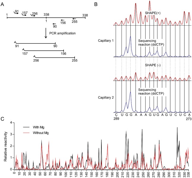
Example of a selective 2′‐hydroxyl acylation analysed by primer extension (SHAPE) experiment adapted to probe the structure of Peach latent mosaic viroid (PLMVd) (+). (A) Representation of the head‐to‐tail dimer of PLMVd and of the primers used to produce the monomeric RNA transcripts of PLMVd starting from different sites. Noncomplementary regions represent the T7 promoter used for the run‐off transcription. (B) The cDNAs obtained from the primer extensions of the SHAPE (+) and SHAPE (–) reactions were electrophoresed on two different capillaries, both of which were accompanied by a sequencing reaction. Both the SHAPE and sequencing reactions were performed with the same primer, except that the fluorophore bound in 5′ was changed (this is represented by the blue and red traces). ddCTP, dideoxycytidine 5′‐triphosphate. (C) The normalized SHAPE reactivities for the reactions performed in either the presence (black) or absence (red) of MgCl2. The results are presented as a function of nucleotide position.