Figure 2.

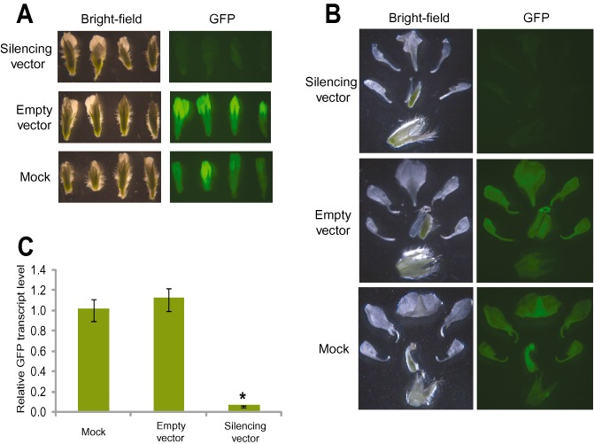
Bean pod mottle virus (BPMV)‐induced gene silencing of the green fluorescent protein transgene (GFP) in floral tissue. (A) GFP silencing in flowers. Flowers at different developmental stages were collected from transgenic GFP soybean plants treated with silencing vector (top panel), BPMV empty vector (middle panel) or mock (bottom panel) and visualized under white light (left panel) and UV light (right panel) and photographed (1‐s exposure on a Zeiss Stemi SV11 stereoscope). Similar results were obtained from three independent experiments. (B) GFP silencing in floral whorls. Hand‐dissected flowers were collected from transgenic GFP soybean plants inoculated with silencing vector (top panel), BPMV empty vector (middle panel) or mock (bottom panel) at 7 weeks post‐virus inoculation and visualized under white light (left panel) and UV light (right panel) and photographed (0.5‐s exposure on a Zeiss Stemi SV11 stereoscope). Note the reduced GFP fluorescence in the flowers and floral whorls collected from the transgenic plants inoculated with silencing construct compared with those obtained from the control treatments. (C) Quantification of gene silencing induced by BPMV in floral tissues using quantitative polymerase chain reaction (qPCR). The mRNA expression levels of GFP were measured in RNA samples isolated from floral tissues of the transgenic GFP plants at 7 weeks post‐virus inoculation and compared with those treated with empty vector or mock. The expression level was calculated using the 2–ΔΔCT method. Data are the average of three biological samples, each with three technical replicates. Mean values significantly different from the mock are indicated by an asterisk as determined by paired t‐test (P < 0.01).
