
Figure 2.
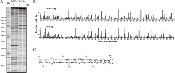
Typical example of the SHAPE probing of the Citrus exocortis viroid clone CEVd.188. (A) Typical SHAPE experiment gel obtained using the primer CEVd AS 258–276 in the primer extension reaction. The lane ddC represents the ladder produced during primer extension using dideoxycytosine. The viroid was probed in either the presence or absence of benzoyl cyanide (BzCN+/–), and either without or with 10 mm Mg2+(–/+). The nucleotide positions on the viroid's structure are indicated on the left of the gel. (B) Histograms generated following analysis of the raw data using the SAFA software after background subtraction and normalization in the absence (top graph) and presence (bottom graph) of magnesium ions. (C) Secondary structure of the region probed in A (from nucleotides 144 to 244) according to SHAPE data incorporated into the RNAstructure software. The region in grey was impossible to analyse as it was located in a poorly resolved region of the gel. The colours are attributed according to the relative SHAPE intensities (black ≤ 0.3, 0.3 < blue < 0.5, 0.5 ≤ orange < 0.7, 0.7 ≤ red).