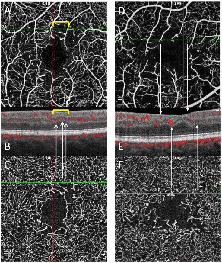
Fig 6. En-face OCTA angiograms and structural B-scans of two T1D patients.
En-face OCTA angiograms of the superficial vascular plexus (SVP, A and D) and deep vascular complex (DVC, C and F) of two T1D patients with the corresponding structural B-scan with angio-overlay (B and E), both passing at the green line shown on the en-face OCTA angiograms. Different patterns of capillary drop-out are seen among the plexuses. In box A, an area of capillary drop-out in the SVP is delimited by the yellow bracket and corresponds to an area of inner retinal thinning and no flow signal in B. In the DVC, the flow signal is visible in the same area as in A, with visible capillaries in both boxes C and B (white arrows indicate red spots on the angio-overlay B-scan). In box D, two arrows from the SVP indicate two bigger vessels with no flow signal between them and a corresponding retinal thinning in E. In the DVC, the flow signal is visible in the same area as in D, with visible capillaries in both boxes F and E (white arrows indicate red spots on the angio-overlay B-scan). The inner nuclear layer and outer plexiform layer show an irregular wavy pattern on both boxes B and E.

