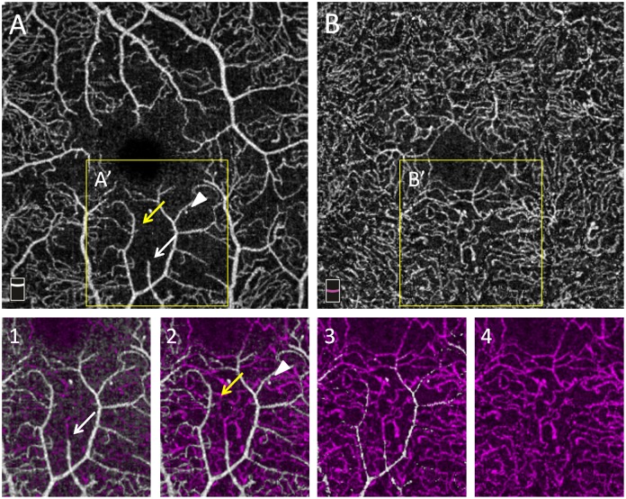
Fig 7. En-face OCTA angiograms of vascular plexuses in a T1D patient.
En-face OCTA angiograms of the superficial vascular plexus (SVP, A) and deep vascular complex (DVC, B) in severe NPDR. A’ and B’ represent the same areas magnified in boxes 1,2,3,4. Boxes 1,2,3,4 represent consecutive color-coded OCTA slabs with the same thickness acquired at increasing depth from the SVP to the DVC. White vessels arise from the SVP while purple vessels arise from the DVC. Arrows (white and yellow) and the white arrow-head indicate residual capillaries in an area of capillary drop-out in the SVP (A) connecting with a denser capillary network in the DVC.

