Figure 4.

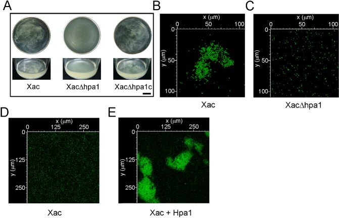
Hpa1 aggregates Xanthomonas axonopodis pv. citri (Xac) bacterial cells. (A) Representative photographs of Xac wild‐type, XacΔhpa1 and XacΔhpa1c strains grown statically in XVM2 medium in 100‐mL flasks. Top panel shows flask top views and bottom panel shows lateral views. Bar, 1 cm. (B) Photograph of a representative aggregate formed by green fluorescent protein (GFP)‐expressing Xac wild‐type previously grown statically in XVM2 medium. (C) Representative photograph of GFP‐expressing XacΔhpa1 grown as in (B). (D, E) Representative photographs of confocal laser scanning microscopy of the GFP‐expressing Xac wild‐type without (D) and with (E) 5 μm Hpa1.
