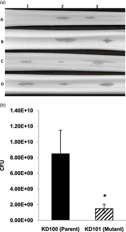
Figure 4.

Celery tissue maceration and bacterial population in planta of Pectobacterium carotovorum strains. (a) Ten microlitres of a bacterial suspension of approximately 3 × 10⁸ colony‐forming units (CFU)/mL was inoculated onto each site of celery petioles. Strains that carried plasmids had chloramphenicol (Cm) added to the suspension. Inoculated celery petioles were placed in a moist container and incubated at 28 °C for approximately 24 h. 1: A, H2O; B, KD100 (CorA+); C, KD101 (CorA‐). 2: A, H2O; B, Ecc71 (CorA+); C, KD102 (CorA‐). 3: A, KD100/pTH19cr (CorA+); B, KD101/pTH19cr (CorA‐); C, KD101/pCKD121 (CorA‐) (CorA+). 4: A, Ecc71/pTH19cr (CorA+); B, KD102/pTH19cr (CorA‐); C, KD102/pCKD121 (CorA‐) (CorA+). (b) Bacterial suspension (10 µL) of approximately 3.5 × 10⁷ CFU/mL was inoculated onto each site of celery petioles. After 20 h of incubation, each inoculated petiole segment of equal size was ground in 25 mm phosphate buffer, diluted and plated. Each bar represents the mean total CFU plus the standard deviation of five replicates. Asterisk indicates significance at P < 0.05.
