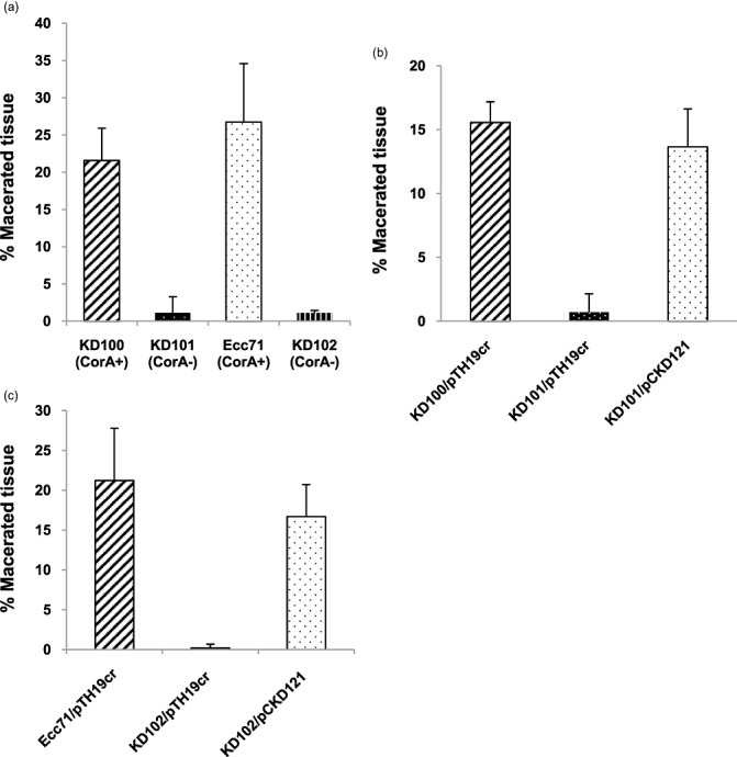
Figure 5.

Carrot tissue maceration by Pectobacterium carotovorum strains. A small well was bored on one side of the carrot disc and inoculated with 50 µL of a bacterial suspension of approximately 7.5 × 10⁷ colony‐forming units (CFU)/mL. Strains that carried plasmids had chloramphenicol (Cm) added to the suspension. Control carrot discs were inoculated with H2O or H2O + Cm. Carrot discs (four replicates each) were placed in a moist container at 28 °C and incubated for 72 h. After 72 h, the amount of tissue maceration was determined (see Experimental procedures). (a) KD100 (CorA+), KD101 (CorA‐), Ecc71 (CorA+) and KD102 (CorA‐); (b) KD100/pTH19cr (CorA+), KD101/pTH19cr (CorA‐) and KD101/pCKD121 (CorA‐) (CorA+); (c) Ecc71/pTH19cr (CorA+), KD102/pTH19cr (CorA‐) and KD102/pCKD121 (CorA‐) (CorA+). The error bars represent the standard deviations.
