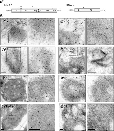
Figure 2.

Viral protein constituents of membranous inclusion structures in Wheat yellow mosaic virus (WYMV)‐infected cells. (A) Schematic presentation of the WYMV genome. Boxes represent the open reading frame of the polyproteins that are cleaved by viral proteases into 10 functional proteins. Black triangles indicate proteinase cleavage sites. (B) Electron micrographs showing immunogold labelling of inclusion structures. Ultrathin sections were prepared from leaves of WYMV‐infected wheat and subjected to immunogold labelling using WYMV P1, P2, P3, NIa‐Pro, VPg, CI, 7K and CP antisera; 10 nm gold particle‐conjugated goat antibody against rabbit immunoglobulin G (IgG) was used as the secondary antiserum. The close‐up views of the dashed rectangular areas are also presented (right images). Bars, 0.2 μm.
