Figure 1.

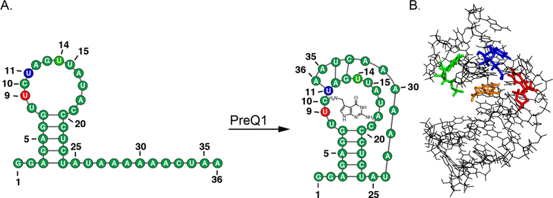
(A) PreQ1-RS riboswitch secondary structure in the unbound and bound state. (B) The bound tertiary structure of PreQ1-RS (PDB 2L1 V) with sites of BFU fluorophore insertion (U9-red, U11-blue, U14-green) and PreQ1 ligand (orange).8

(A) PreQ1-RS riboswitch secondary structure in the unbound and bound state. (B) The bound tertiary structure of PreQ1-RS (PDB 2L1 V) with sites of BFU fluorophore insertion (U9-red, U11-blue, U14-green) and PreQ1 ligand (orange).8