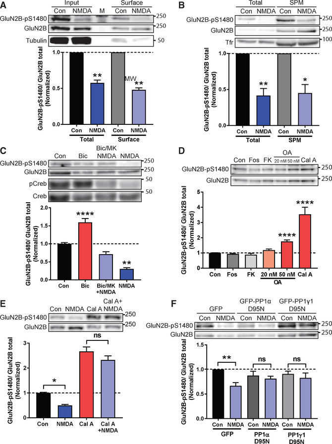
Figure 2. Co-activation of Synaptic and Extrasynaptic NMDARs Promotes PP1-Mediated Dephosphorylation of S1480 on Surface-Expressed GluN2B.
Levels of normalized GluN2B-pS1480 were analyzed by immunoblotting from cortical primary neurons after the indicated manipulations. Graphs represent mean ± SEM. *p < 0.05; **p < 0.01; ****p < 0.0001 using a Mann-Whitney U test (A and B) or Kruskal-Wallis H test (C-F).
(A) Biotinylation experiment in DIV24-DIV28 neurons after induction of GluN2B-pS1480 dephosphorylation by NMDA incubation (50 μM for 10 min). Tubulin is shown as a control. M denotes marker. n = 5.
(B) Isolation of the synaptic plasma membrane (SPM) fraction from DIV14-DIV21 neurons following NMDA treatment as before. Transferrin receptor (Tfr) is shown as a control. n = 5.
(C) Synaptic, extrasynaptic, or global activation of NMDARs in DIV21-DIV28 neurons. pCreb and Creb are shown as controls. n = 3.
(D) Incubation of DIV14-DIV17 neurons with phosphatase inhibitors for 45 min: fostriecin for PP2A/4 (Fos; 1 μM); FK506 for PP2B (FK; 1 μM); okadaic acid for PP2A at 20 nM and PP2A/PP1 at 50 nM (OA; 20 or 50 nM); and calyculin A for PP1/PP2A (Cal A; 100 nM). n = 10.
(E) Pre-incubation of DIV14-DIV21 neurons with 100 nM Cal A for 45 min before NMDA treatment as before. p = 0.66 (Cal A versus Cal A + NMDA). n = 14.
(F) Lentiviral transduction of the dominant negative forms of GFP-PP1 α and γ1 (PP1α/γ1 D95N) and GFP (as a control) in DIV14-DIV17 neurons for 7–10 days. GluN2B-pS1480 dephosphorylation was induced with NMDA as before. p > 0.99 (PP1α) and p = 0.78 (PP1γ1). n = 6.
See also Figures S1 and S2.

