. 2019 Jul 10;8:e44752. doi: 10.7554/eLife.44752

Figure 4. RNAs targeted to protrusions are similarly translated in both internal and peripheral locations.

(A) Live imaging snapshots of cells expressing the indicated translation reporters. GFP/mCherry intensity of individual spots (indicating translation efficiency) was plotted as a function of distance from the cell edge. More than 200 particles were analyzed from approximately 20 cells. Best fit curves with 95% confidence intervals are overlaid on the graphs. Scale bars: 5 μm. (B) Cumulative frequency distribution plot of translation reporter particles (from panel A) with increasing distance from the cell edge.

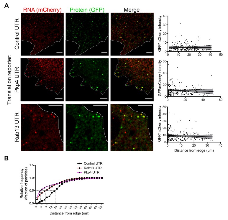
Figure 4.

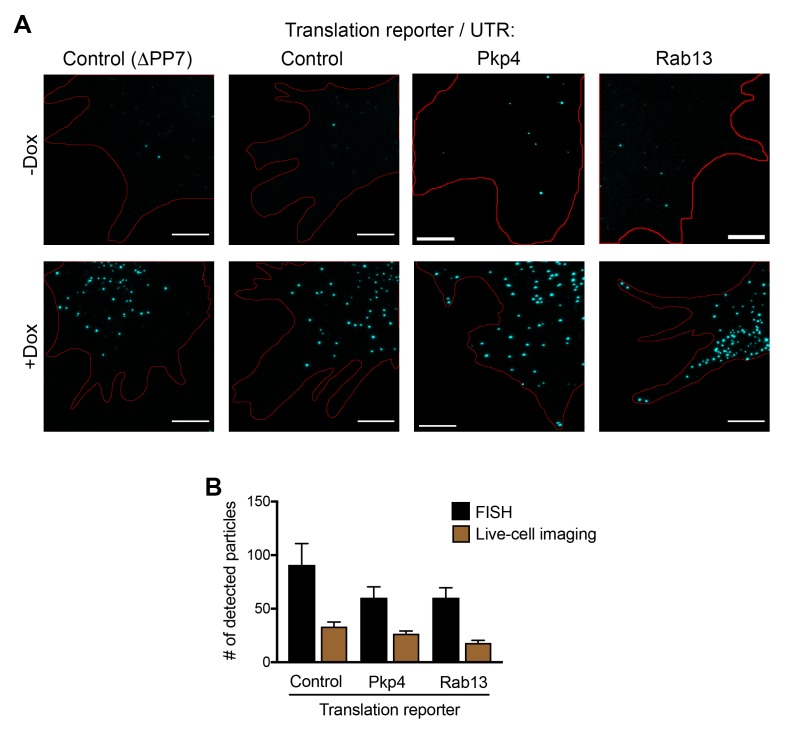
Figure 4—figure supplement 1. Expression levels of translation reporters and comparison with live-cell imaging.

Figure 4—figure supplement 1.

(A) In situ hybridization of cells expressing the indicated translation reporters in the absence or presence of doxycycline. Scale bars: 5 μm. Red line:cell outline. (B) Comparison of the number of translation reporter particles detected per cell, after doxycycline addition, by in situ hybridization or live-cell imaging. Expression levels, assessed by FISH, are not significantly higher than endogenous APC-dependent RNAs (compare to values presented in Figure 5—figure supplement 2). Fewer particles are detected by live-cell imaging likely indicating that fast-moving particles cannot be resolved with our current acquisition methods.
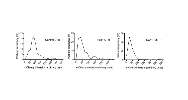
Figure 4—figure supplement 2. Intensity histograms of translation reporter particles.

Figure 4—figure supplement 2.

Frequency distribution plots of mCherry intensities exhibited by particles of the indicated translation reporters. The existence of a single major peak suggests that the majority of observed particles are single molecules.
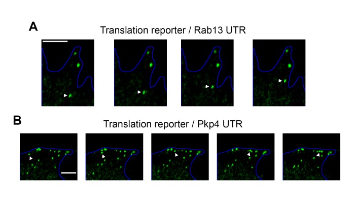
Figure 4—figure supplement 3. Examples of directionally persistent particles.

Figure 4—figure supplement 3.

(A, B) Snapshots of time lapse imaging of cells expressing the indicated localized translation reporters. The GFP channel was recorded showing translated RNAs. Arrowheads point to particles that move in a directionally persistent manner during the time of observation. Scale bars: 3 μm. Full length movies of these samples are presented in Video 5 (A) and Video 6 (B).