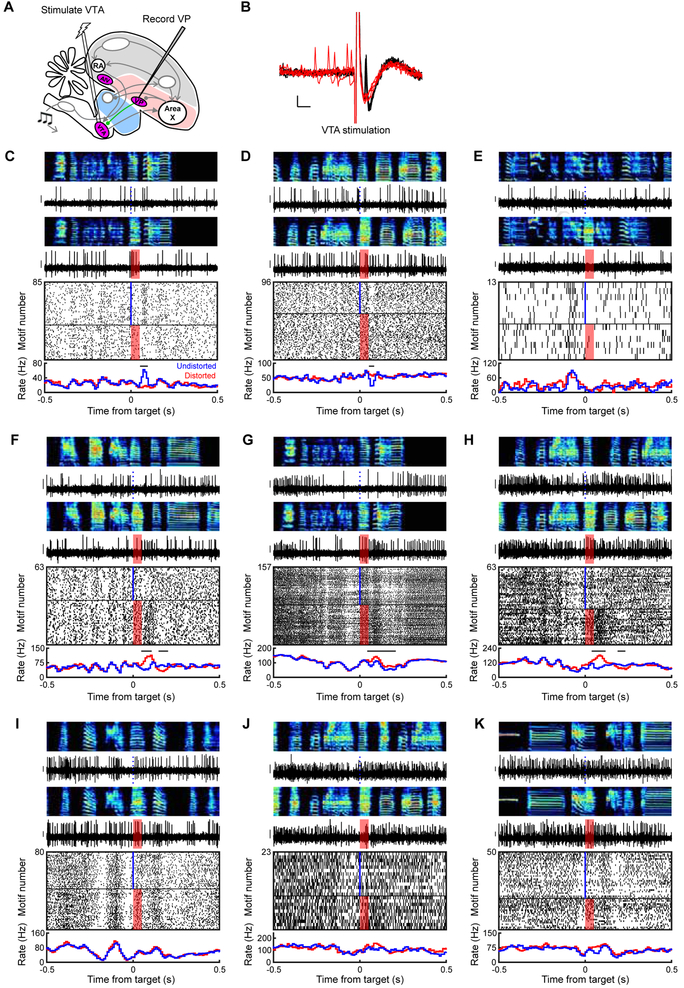
Figure 5. VP sends diverse error- and prediction-related signals to VTA. See also Figure S3.
(A) Stimulation and recording electrodes were chronically implanted into VTA and VP, respectively, for antidromic identification of VTA-projecting VP neurons (VPVTA). (B) Antidromic (black) and collision (red) testing of the neuron shown in (C). Scale bars: 1ms (horizontal) and 0.1mV (vertical). (C to K) Song-locked firing patterns of 9 VPVTA neurons, plotted as in Figure 3B, reveal diverse responses including prediction errors (C and D), pre-target burst (E), error-induced activation (F-H), and pre-target pauses (F to J). Y scale bar for spiking activity is 0.15 mV.

