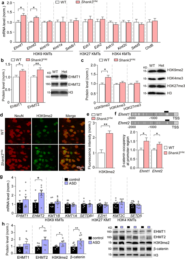
Figure 1. EHMT1/2 and H3K9me2 levels are specifically elevated in the PFC of Shank3+/ΔC mice and autistic human patients.
(a) Quantitative real-time RT-PCR data on the mRNA level of 10 histone methyltransferases catalyzing H3K9, H3K27 or H3K4 methylation, and Chd8 in the frontal cortical tissue from WT and Shank3+/ΔC mice (Ehmt1/2: n = 13–16/group; others: n = 6–8/group, * P < 0.05, t-test). (b, c) Quantitation and representative immunoblots of EHMT1 and EHMT2 protein levels (b) and H3K9me2, H3K4me3 and H3K27me3 levels (c) in the nuclear fraction of prefrontal cortical tissue from WT and Shank3+/ΔC mice (n = 9–10/group, * P < 0.05, ** P < 0.01, t-test). (d) Representative confocal images of immunohistochemical staining of H3K9me2 and NeuN in PFC of WT and Shank3+/ΔC mice. (e) Plots showing the level of H3K9me2 in PFC of WT and Shank3+/ΔC mice. (n = 15–30 slices/5 animals; *** P < 0.001, t-test). (f) ChIP assay data showing β-catenin binding at Ehmt1 and Ehmt2 promoter regions in PFC lysates from WT vs. Shank3+/ΔC mice (n = 7–8/group, * P < 0.05, t-test). Top: schematic graph showing the location of primer covering the TCF-LEF binding motif (labeled with vertical gray lines). (g) qPCR data on the mRNA level of EHMT1, EHMT2 and several other histone methyltransferases catalyzing H3K9, H3K27 or H3K4 methylation in Brodmann’s Area 9 (BA9) of postmortem tissue from autistic human patients and healthy controls (n = 7–10/group, * P < 0.05, t-test). (h) Quantitation and representative immunoblots of EHMT1, EHMT2, H3K9me2 and β-catenin levels in the nuclear fraction of BA9 from autistic human patients and healthy controls (n = 6–9/group, * P < 0.05, ** P < 0.01, t-test). Data are expressed as mean ± sem.

