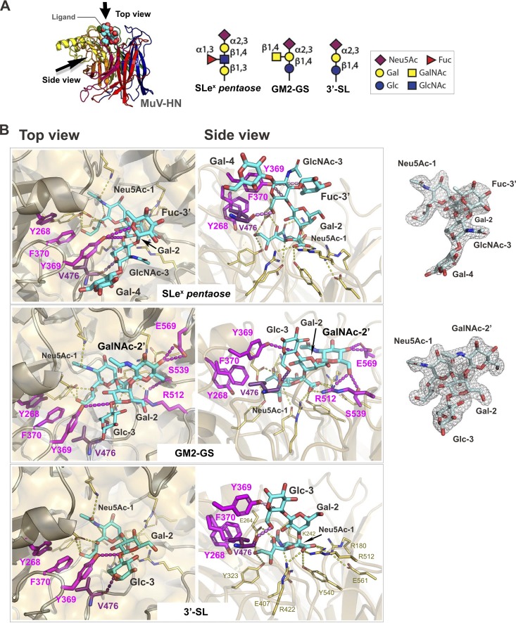
FIG 2.
Interactions between the glycans and MuV-HN protein. (A) Overall structure of the MuV-HN−SLeX pentaose complex structure (left) and schematic of the glycan receptor derivatives for MuV (right). (B) Binding site of SLeX pentaose, GM2-GS, and 3′-SL in the MuV-HN−glycan complex structures. SLeX pentaose, GM2-GS, and 3′-SL are shown in cyan. Top (left) and side (middle) views are shown. The omit Fo – Fc (the observed and calculated structure factor amplitude, respectively) maps (3.0σ) for SLeX pentaose and GM2-GS are also shown (right). The MuV-HN residues directly involved in the interaction with Sia-1 are shown in yellow, whereas the ones interacting with the non-Sia-1 moieties of the glycans are indicated by magenta. Nitrogen atom, blue; oxygen atom, red.

