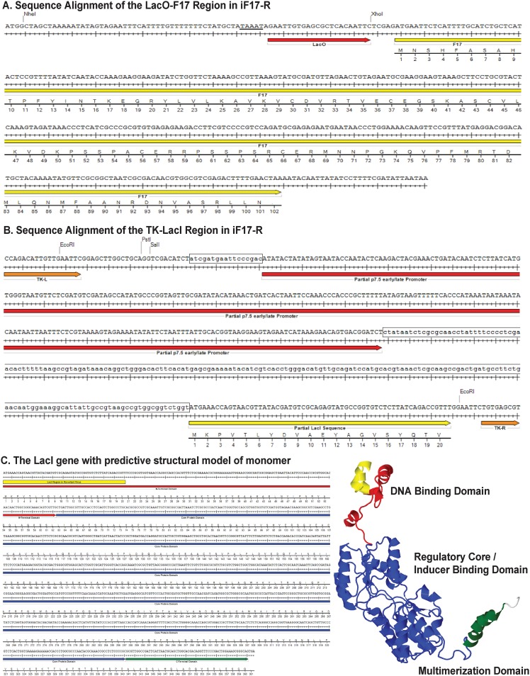
FIG 7.
Map of mutations detected in the iF17-R stock. (A) Sequence alignment for LacO gene repressor binding site (red arrow) and F17 open reading frame (yellow arrow). Canonical TAAAT motif in the late promoter is underlined. (B) The partial sequences of the p7.5 early/late promoter (red arrow) and LacI gene repressor (yellow arrow) flanked by left and right TK sequences (orange arrows) at the insertion site. Restriction sites used in the original generation of the vector are shown. Boxed regions highlight sequence divergence detected in the iF17-R stock at the p7.5 promoter or loss of large portions of the LacI gene open reading frame. (C) Sequence alignment and predicted structure of the LacI gene repressor. Red is the N-terminal DNA binding domain, blue is the core regulatory region, green is the C-terminal tetramerization domain, and yellow represents the partial DNA binding domain that remains in the iF17-R virus.

