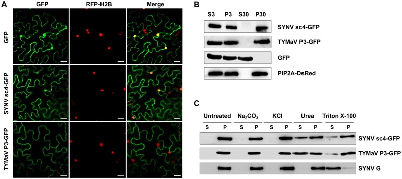
FIG 3.
Subcellular localization and microsomal association analyses of SYNV sc4-GFP and TYMaV P3-GFP. (A) Confocal micrographs of leaf epidermal cells of transgenic N. benthamiana plants expressing RFP fused to histone 2B (RFP-H2B) that had been infiltrated to express GFP, SYNV sc4-GFP, or TYMaV P3-GFP. Scale bars = 20 μm. (B) Protein extracts from N. benthamiana plants expressing either SYNV sc4-GFP, TYMaV P3-GFP, or the transmembrane SYNV G protein, together with GFP and PIP2A-DsRed marker proteins, were separated into supernatant (S3) and pellet (P3) fractions by centrifugation at 3,000 × g for 10 min, and the S3 fraction was further separated into the pellet (P30) and supernatant (S30) fractions by ultracentrifugation at 30,000 × g for 1 h. Protein gel blots revealed that SYNV sc4 and TYMaV P3 were present only in the P30 fraction. (C) The P30 fractions in (B) were divided into several aliquots and treated with either 0.1 M Na2CO3 (pH 11.5), 1 M KCl, 7 M urea, or 1% Triton X-100. Samples were again separated into S and P fractions by ultracentrifugation and subjected to protein gel blot analyses with anti-GFP antibodies or anti-G antibodies.

