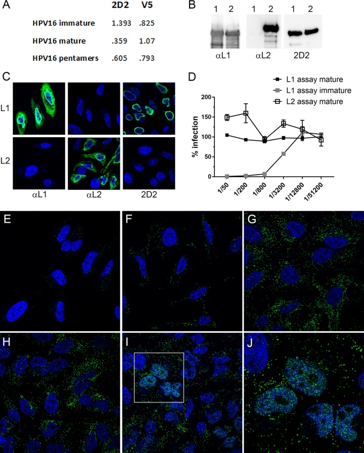
FIG 1.
Characterization of 2D2 mAb. (A) Differential ELISA reactivities of 2D2 to HPV16 immature and mature L1/L2 capsids and pentamers. Capture ELISAs were performed by coating plates with a capture reagent, rabbit anti-HPV16 VLP. HPV16 preparations were applied at 150 ng/well. 2D2 or V5 antibody (anti-HPV16 L1 VLP) was added at a dilution of 1:10 (hybridoma supernatant) or 1:5,000 (ascites), respectively. Bound antibody was detected with horseradish peroxidase-conjugated secondary antibody. (B) Detection of L1 and L2 by Western blotting. In each instance, L1-only VLPs were run in lane 1, and L1 and L2 PsV was run in lane 2. Proteins were detected on the three blots with anti-HPV16 L1, anti-HPV16 L2, or 2D2. (C) HeLa cells that were transfected with either an L1 or an L2 expression plasmid were immunostained for the same antigens. (D) Neutralization of immature and mature PsVs with 2D2. Neutralization assays were performed in triplicate across a 4-fold dilution series of the 2D2 hybridoma supernatant starting at a 1:50 dilution. In the L1-based assay, antibody dilutions are applied to PsV prior to addition to 293TT cells, whereas in the L2-based assay, capsids are applied to an epithelial cell extracellular matrix and cleaved with furin prior to exposure of the capsids to the antibodies and target cells. (E to I) Time course of PsV detection. HeLa cells were either uninfected (E) or infected with HPV16 PsV for 1 h (F), 2 h (G), 7 h (H), or 24 h (I). (J) Magnification of the inset in panel I. L1 was stained with 2D2 and donkey anti-mouse IgG-Alexa Fluor 488 (green channel). Nuclei are stained with DAPI (4′,6-diamidino-2-phenylindole) (blue channel). This temporal exposure of the 2D2 epitope was observed in multiple time course experiments.

