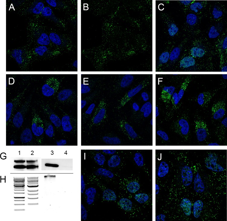
FIG 2.
L1 nuclear delivery is L2 dependent and prevented with biochemical inhibition of infection. (A to F) Either HPV16 L1-only VLPs (A and B) or L1/L2 PsV (C to F) was applied to HeLa cells for 24 h and stained with 2D2 and donkey anti-mouse IgG-Alexa Fluor 488.(A and B) Duplicate images with the DAPI nuclear signal removed from the second panel to better visualize L1 nuclear localization. (C to F) The following inhibitors were added during PsV incubation: 10 μg cycloheximide (C), 10 μM RVKR-cmk (furin inhibitor) (D), 300 nM compound XXI (γ-secretase inhibitor) (E), and 10 μM cyclosporine (cyclophilin inhibitor) (F). (G to J) Nuclear L1 delivery does not require packaged DNA. Particles were confirmed to lack DNA by Western blot determination of histone H3 content. (G) Western blots of L1 (bottom bands) and L2 (top bands). Lane 1, full particles; lane 2, empty particles; lanes 3 and 4, Western blots for histone H3 in the same sample order. (H) DNA content extracted from these particles. DNA ladders are shown in the first two lanes, the third lane shows DNA extracted from full particles, and the fourth lane shows the extraction from the empty particles. (I and J) Cells that were infected in parallel with either full particles (I) or empty particles (J), stained with antibody H16.2F at 24 h postinfection. Nuclei are stained with DAPI.

