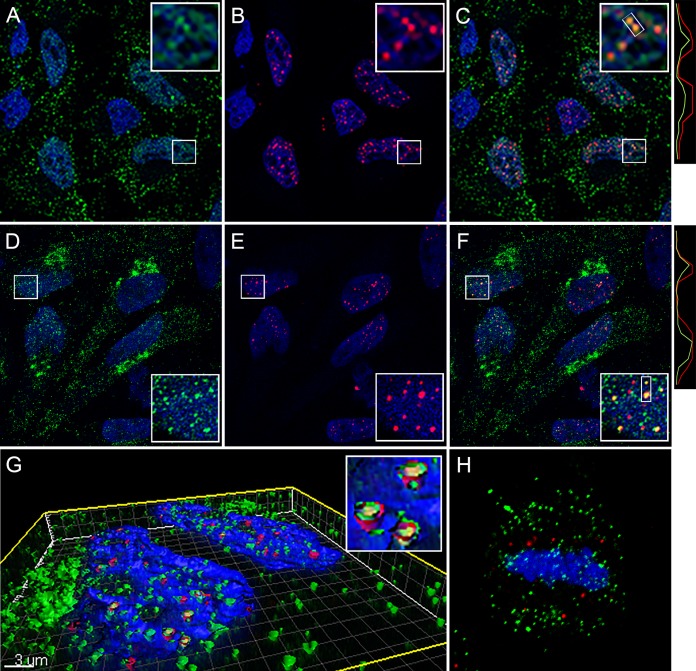
FIG 3.
Nuclear localization of L1 at ND10. (A to C) Colocalization of 2D2 staining with PML at 24 h postinfection. (A) L1 was detected with 2D2 and donkey anti-mouse IgG-Alexa Fluor 488 (green). (B) PML was detected with a rabbit polyclonal antiserum and donkey anti-rabbit IgG-Alexa Fluor 594 (red). (C) Merged signals. Histograms illustrating the degree of colocalization through the indicated region are on the right. (D to F) The same staining in cells that were extracted with 1% NP-40 prior to fixation. (G) A Z-stack series from cells that underwent this treatment was surface rendered. (H) Detection of L1 on mitotic chromosomes in NP-40-extracted cells. Nuclei are stained with DAPI in all panels.

