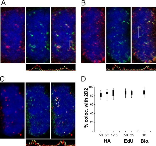
FIG 4.
Colocalization of 2D2-reactive L1 with L2, vDNA, and biotinylated pseudovirions on mitotic chromosomes. All panels show mitotic chromosomes from HeLa cell cultures at 24 h postinfection. (A) Colocalization of 2D2 staining with L2 in a single slice from a Z-stack series. 2D2 binding was detected with donkey anti-mouse IgG-Alexa Fluor 488 (green). HA-tagged L2 was detected with a goat polyclonal antiserum and donkey anti-goat IgG-Alexa Fluor 594 (red). (B) Colocalization of 2D2 staining with the vDNA in a single slice from a Z-stack series. HPV16 PsV containing EdU-labeled DNA is shown. L1 was stained with 2D2 and donkey anti-mouse-Alexa Fluor 594 (red). Cells were then processed for EdU detection with Click-iT Alexa Fluor 488 reagents (green). (C) Colocalization of 2D2 staining (red) with biotinylated HPV16 PsV particles, detected with Alexa 488-coupled streptavidin. Histograms illustrating the degree of colocalization through the indicated regions are shown below the corresponding images. (D) Percent colocalization between 2D2-stained puncta and HA, EdU, or biotin across a range of input concentrations, as indicated. The microscopic images shown correspond to 25 ng input PsV for L2 and EdU colocalization and 10 ng for colocalization with biotinylated capsids.

