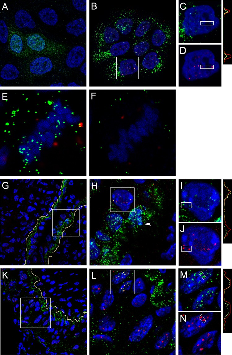
FIG 6.
Nuclear L1 detected in keratinocytes. (A to D) HaCaT cells were infected with HPV16 PsV for 24 h and processed for IF with either 2D2 (A) or human monoclonal anti-L1 mAb 7nv15 (B and C) using either donkey anti-mouse IgG-Alexa Fluor 488 or donkey anti-human IgG-Alexa Fluor 488 secondary antibody conjugates, respectively. 7nv15 staining was performed in conjunction with PML protein detection (B and D) with rabbit anti-PML and donkey anti-rabbit IgG-Alexa Fluor 594. Colocalization through the indicated region is illustrated in the histogram. (E and F) Differential permeabilization staining. Shown is the accessibility of 7nv15 antibody to L1 on the mitotic chromosomes on HaCaT cells following NP-40 permeabilization (E) or digitonin permeabilization (F) followed by the secondary antibody donkey anti-mouse-Alexa Fluor 488. PML was detected with a rabbit antiserum and donkey anti-rabbit-Alexa Fluor 594. A central section obtained from a Z-stack series is shown for each. (G to N) In vivo staining. The presence of nuclear L1 following murine intravaginal infection was determined by staining tissues with 7nv15 and anti-PML. Tissues were prepared at 18 h (G to J) or 48 h (K to N) postinstillation. The boxed regions are magnified in the adjacent panels. The split panels show 7nv15 anti-L1 staining in green (I and M) or anti-PML staining in red (J and N). Nuclei are stained with DAPI (blue). Histograms illustrating the degree of colocalization through the indicated region are on the right. The basement membrane is traced in yellow in the lower-magnification images.

