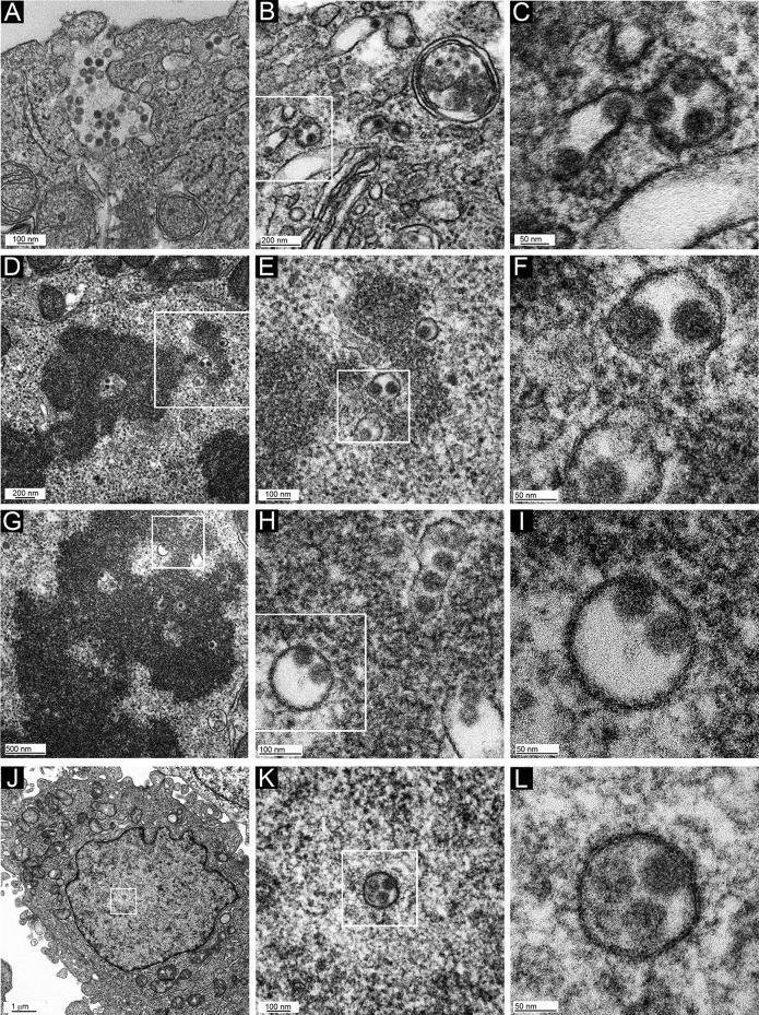
FIG 7.
TEM micrographs of HPV16-infected HeLa cells. Cells were infected for 24 h with HPV16 PsV and processed for TEM imaging. Panel inset boxes show the area magnified in the following panel. Two different mitotic cells were chosen for panels D to F and G to I. Cells shown in panels A to C and J to L are in interphase.

