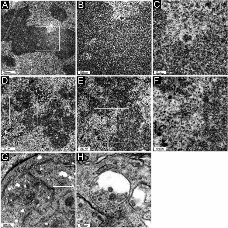
FIG 8.
TEM micrographs of control HeLa cells. Mitotic cells (A to F) and an interphase cell (G and H) are shown. (A to C) Uninfected cells; (D to H) cells incubated with L1-only VLPs for 24 h. Panel inset boxes show the area magnified in following panel. Note that there is some electron-dense material in the chromosomal region, but it is irregular in shape and not within vesicles. This is most apparent in panels B and C. Panel G and the magnification in panel H show the localization of L1-only particles within endosomal vesicles.

