FIG 6.
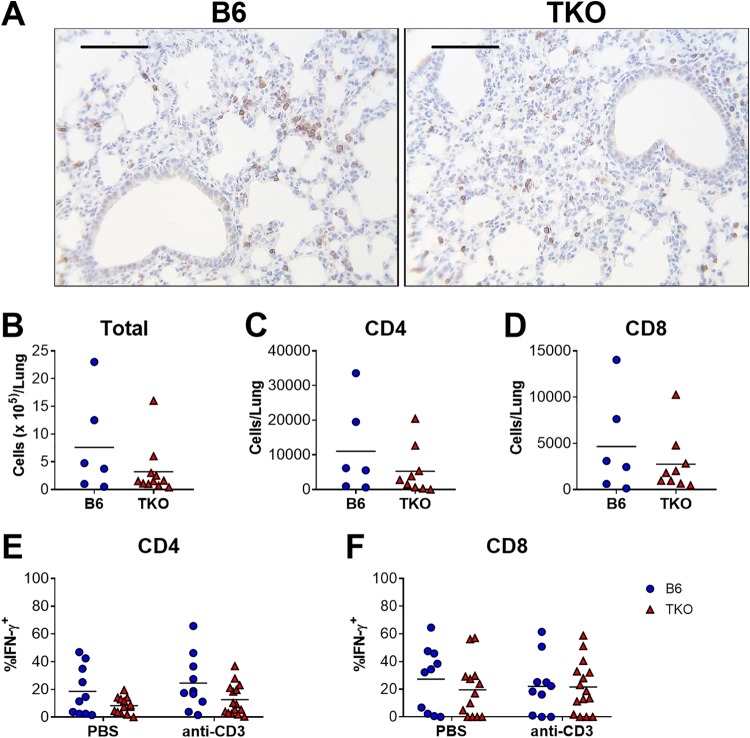
Effects of immunoproteasome deficiency on T cell responses to MAV-1 infection in neonatal mice. B6 and TKO mice were infected i.n. with MAV-1 at 7 days of age. (A) Sections from lungs of infected mice harvested at 9 dpi were stained with antibody specific for CD3. CD3+ cells are stained dark brown. Representative images from n = 3 mice per group are presented. Scale bars, 100 μm. (B to F) Lung leukocytes were isolated from lungs of infected mice at 7 dpi. (B) Total cell numbers were quantified with a hemocytometer. Cells were stimulated overnight with anti-CD3 antibody or left unstimulated (PBS controls). Overall numbers of CD4+ (C) and CD8+ (D) T cells in lungs were calculated based on total lung cell counts and the percentage of CD4+ or CD8+ cells in unstimulated fractions, as determined by flow cytometry (n = 6 to 9 per group). Intracellular cytokine staining was used to quantify the percentages of CD4+ (E) and CD8+ (F) cells that were IFN-γ+ (n = 10 to 15 per group). Statistical comparisons were made using the Mann-Whitney test (B to D) or two-way ANOVA (E and F), followed by Bonferroni’s multiple-comparison tests. No statistically significant differences were detected.

