FIG 1.
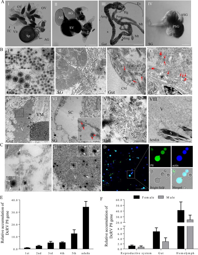
DcRV particles are widely distributed throughout the organs of D. citri. (A) Transmitted light micrograph of dissected organs of D. citri. The organs mainly consist of the reproductive system (I and II), gut (III), and salivary gland (IV). Bars are 200 μm (I, II, and III) and 50 μm (IV). (B) Electron micrographs showing DcRV virions aggregated into crystalline arrays, scattered throughout the cytoplasm, distributed at the periphery viroplasm, or contained in vesicular compartments in different D. citri organs. Insets are enlarged images of the boxed areas in each panel. Arrows indicate the free virions in the tissues. Bars are 200 nm (I), 700 nm (II, VII, and VIII), 500 nm (III and IV), 1 μm (V), and 3 μm (27). OV, ovary; LO, lateral oviduct; Sp, spermatheca; AG, accessory gland; TC, trophic chamber; Vit, vitellarium; T, testis; SV, seminal vesicle; Es, esophagus; FC, filter chamber; Amg, anterior midgut; Mmg, middle midgut; Pmg, posterior midgut; Hg, hind gut; Mt, Malpighian tubule; SG, salivary gland; PSG, principle salivary gland; ASG, accessory salivary gland; CM, circular muscle; EC, epithelial cell; V, viroplasm; VM, visceral muscle; SC, salivary cavity; S, sperm. (C) Immunogold labeling of P8 on outer shells of virions in the cytoplasm (I) and on the viroplasm (II) of epithelial cells of guts. White arrows mark gold particles. Insets i, ii, and iii are enlarged images of the boxed areas of panels I and II. Bars, 100 nm. (D) Immunofluorescence microscopy showing that DcRV is distributed in the hemolymph of DcRV-infected D. citri. Hemocytes were immunolabeled with P8-FITC antibody (green) and actin dye phalloidin-Alexa Fluor 647 carboxylic acid (blue) and presented single sections examined by confocal microscopy. Bars, 50 μm. (E) RT-qPCR showing that DcRV accumulation gradually increased with the development of D. citri. (F) RT-qPCR showing gender preference and tissue-specific distribution of DcRV. The results were normalized against the level of the actin mRNA, and the gene accumulation levels of P8 in 1st-instar nymphs (E) or in reproductive systems of males (F) was normalized as 1. Means (±standard deviations [SD]) from 3 biological replicates are shown.

