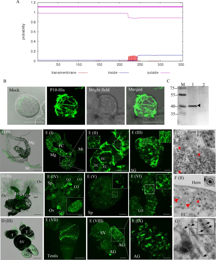
FIG 3.
DcRV P10 is associated with tubules that can package virions in DcRV-infected D. citri. (A) Prediction of hydrophobic regions for DcRV P10 generated with TMHMM. (B) Expression of P10 fused with a His tag showing filament-like structures in recombinant baculovirus-infected Sf9 cells at 72 hpi. Sf9 cells were treated with fluorescein isothiocyanate-conjugated His-6×His tag antibody. The image of mock infection with green fluorescence (His-FITC antibody) was merged under a background of transmitted light. Bar, 10 μm. (C) Western blot analyses showing the specificity of antibodies against the P10 peptide. Total proteins of Californian DcRV-infected and uninfected D. citri insects were separated by SDS-PAGE and analyzed with P10 antibodies. Lane M, protein ladder; lane 1, protein extracts from DcRV-infected D. citri; lane 2, protein extracts from uninfected D. citri. (D) Immunofluorescence microscopy showing the absence of P10 in the alimentary canal (I), female reproductive system (II), and male reproductive system (III) of uninfected D. citri. The images with green fluorescence (P10-FITC antibody) were merged under a background of transmitted light. Bars, 200 μm. (E) Immunofluorescence microscopy showing that P10 widely distributes in the body of DcRV-infected California D. citri. The internal organs of DcRV-infected D. citri were treated with P10-FITC antibody (green) and examined by confocal microscopy. Images are presented in stacked sections. Insets are enlarged images of the boxed areas in each panel. Bars are 200 μm (I), 100 μm (II, IV, VII, and VIII), 20 μm (VI and IX), and 50 μm (III and V). (F) Electron micrographs showing virion-containing tubules in the cytoplasm of epithelial cells of guts. Insets are enlarged images of the boxed areas. Red arrows or arrowheads indicate the longitudinal or transverse sections of tubules packaging DcRV virions. Bars, 200 μm. (G) Immunogold labeling of P10 on virion-containing tubules in the cytoplasm of epithelial cells of guts. Black arrows mark gold particles. The inset is an enlarged image of the boxed area. Bars, 200 μm. Images are representative of the results of more than 3 experiments. FC, filter chamber; Mg, midgut; Mt, Malpighian tubule; EC, epithelial cell; SG, salivary gland; Ov, ovary; Sp, spermatheca; LO, lateral oviduct; CO, common oviduct; AG, accessory gland; SV, seminal vesicle; Hem, hemolymph.

