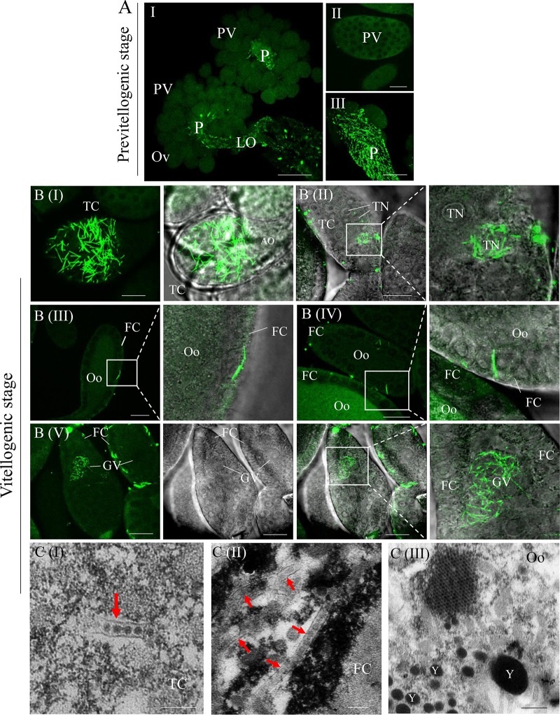
FIG 4.
P10 tubules carry DcRV virions into the ovary at the vitellogenic stage. (A) Immunofluorescence microscopy showing that P10 tubules fail to localize to the ovarioles at the previtellogenic stage. Bars are 100 μm (I), 20 μm (II), and 50 μm (III). (B) Immunofluorescence microscopy showing the distribution of P10 tubules in the trophic chamber and oocyte-developed ovarioles at the vitellogenic stage. The internal ovaries of DcRV-infected D. citri insects were immunolabeled with P10-FITC (green) and then examined by confocal microscopy. Images are presented in stacked sections. The enlarged images display green fluorescence (P10-FITC) and bright fields of the merged images of the boxed areas in each panel, indicating the P10 distribution in the ovary. Bar, 20 μm. (C) Electron micrographs showing virion-containing tubules (arrows) in the vitellogenic ovary. Insets are enlarged images of the boxed areas in each panel. Bars are 200 nm (I and II) and 400 nm (III). Ov, ovary; PV, previtellogenic ovariole; P, pedicle; LO, lateral oviduct; TC, trophic chamber; AO, arrested oocyte; TN, trophocyte nuclei; FC, follicular cell; Oo, oocytes; GV, germinal vesicle; Y, yolk. All micrographs are representative of at least 3 replicates.

