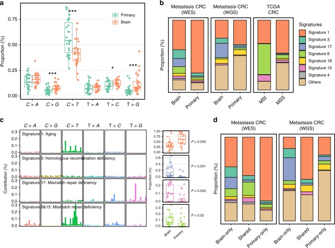
Fig. 1.
Divergent mutational features in brain metastases and matched primary tumors of CRC patients. a Six mutational subtypes in BM and primary tissues. The BM tissues are presented in orange, and the primary tissues are presented in cyan. b Major mutational signatures in our patients and TCGA CRC patients. We reconstructed the proportion of mutational signatures of each sample based on a predefined mutational spectrum of 30 COSMIC signatures. The proportion of the top seven signatures in BM is presented. c HRD and MMRD signatures were significantly elevated in BM tissues compared with those in primary tissues. Four mutational signatures with a significant difference between BM and primary tissues are shown in the figure. The mutation characteristics are presented at the left side and the box plot of the proportion of each signature is presented at the right side. The box plot displays the first and third quartiles (top and bottom of the boxes), median (band inside the boxes), and lowest and highest point within 1.5 times the interquartile range of the lower and higher quartile (whiskers). d All mutations were classified into three groups—brain only, shared and primary only—and mutational signatures were extracted in each group

