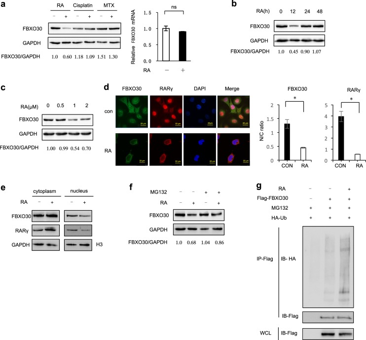
Fig. 4. RA treatment promotes to ubiquitylate FBXO30.
a HEK293 cells were treated with RA (1 μM, 12 h) or Cispaltin (100 μmol/L) or MTX (1 μM) for the indicated times. Cell lysates were harvested to detect FBXO30 level (left). Real-time PCR analysis showed no effect of RA on FBXO30 mRNA level (right). Mean values and SD are depicted. b HEK293 cells were treated with RA (1 μM) for the indicated time. Cell lysates were harvested to detect FBXO30 level. c HEK293 cells were treated with RA 12 h for the indicated concentration. Cell lysates were harvested to detect FBXO30 level. d HEK293 cells were treated with RA (1 μM) for 12 h. Direct visualization or indirect immunofluorescence was performed. Scale bars, 22 μm. e HEK293 cells were treated with RA (1 μM) for 12 h. Distribution of endogenous FBXO30 in HEK293 cells was determined by cell fractionation. The fractions were subjected to western blot with the indicated antibodies. H3 is a nucleolar marker protein. f HEK293 cells were treated with RA (1 μM) and/or MG132 for 12 h. Aliquots of total lysates were immunoblotted to detect anti-FBXO30 antibody. g HEK293 cells transfected with HA-Ub and FBXO30 were treated with MG132. After RA treatment, the ubiquitylation of FBXO30 was analyzed

