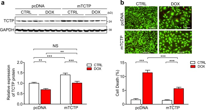
Fig. 5. Exogenous supplementation of TCTP rescued DOX-induced cardiomyocyte death.
H9C2 cells were transfected with pcDNA3.1-control vector or pcDNA3.1 mouse TCTP expression plasmid (mTCTP) for 24 h and then treated with DOX (0.1 μM) or vehicle for 48 h. a Protein expression of TCTP and GAPDH was analyzed by western blotting (n = 8). b Cell death was determined by calcein-AM/ethidium homodimer-1 staining (n = 4). Live and dead cells were distinguished by calcein-AM (green) and ethidium homodimer-1 (red) staining, respectively. Scale bar, 200 μm. *P < 0.05, **P < 0.01, ***P < 0.001. Two-way ANOVA followed by Bonferroni’s test

