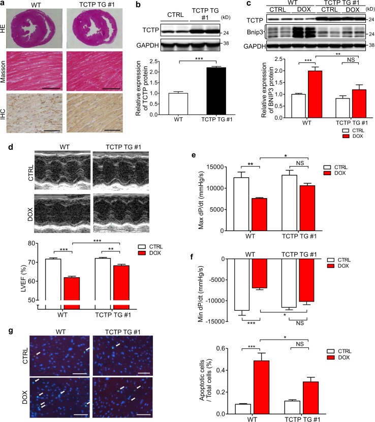
Fig. 6. Cardiomyocyte-specific TCTP overexpression protected mice against DOX-induced cardiac dysfunction.
a Representative images of sections from WT and TCTP TG mouse hearts stained with hematoxylin and eosin (HE) (top) and Masson trichrome (middle). Immunohistochemistry (IHC) with TCTP antibody in heart tissues of WT and TCTP TG mice (bottom). Scale bar, 50 μm. b TCTP and GAPDH protein expression in the left ventricle of WT and TCTP TG #1 mice (n = 5). c WT and TCTP TG mice were treated intraperitoneally with DOX (3 mg/kg) or normal saline (CTRL). DOX was administered three times a week, with a total dose of 24 mg/kg. Protein expression levels of TCTP, Bnip3, and GAPDH in the hearts were analyzed 24 h after the treatment (n = 4–5). d–g five weeks after the first injection, LVEF was evaluated by echocardiography (d) (n = 6–9). Max dP/dt (e) and Min dP/dt (f) were evaluated by cardiac catheterization (n = 6–7). g Cardiac apoptosis in the myocardium was assessed by TUNEL assay in heart sections from WT and TCTP TG mice with DOX or normal saline (CTRL) treatment. Nuclei were stained with DAPI (blue). Representative images are shown to the left. The white arrows indicate apoptotic cells in the sections (n = 4–7). Scale bar, 50 μm. *P < 0.05, **P < 0.01, ***P < 0.001. Unpaired, two-tailed Student’s t-test (b) or two-way ANOVA followed by Bonferroni’s test (c–g)

