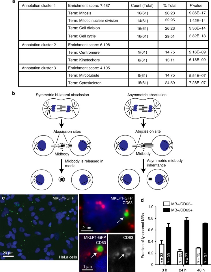
Fig. 1.
Post-abscission MBs are internalized and accumulate in interphase cells. a DAVID functional cluster analysis of mRNAs up-regulated in GFP-MB+ cells. P-value is represented by modified Fisher exact P-value. b Schematic representation of asymmetric and symmetric cell abscission. c, d HeLa cells were incubated with purified GFP-MBs for 3 h. Cells were then washed and incubated for additional 3 h before fixation and staining with anti-CD63 antibodies. In c arrow in top inset shows MB that does not colocalize with CD63, while arrow in bottom insets shows MB that is surrounded by CD63-positive membrane. Panel d shows quantification of the colocalization between GFP-MBs and CD63. n is the number of internalized MBs counted for each condition. Three biological replicates were used to obtain data

