Fig. 4.
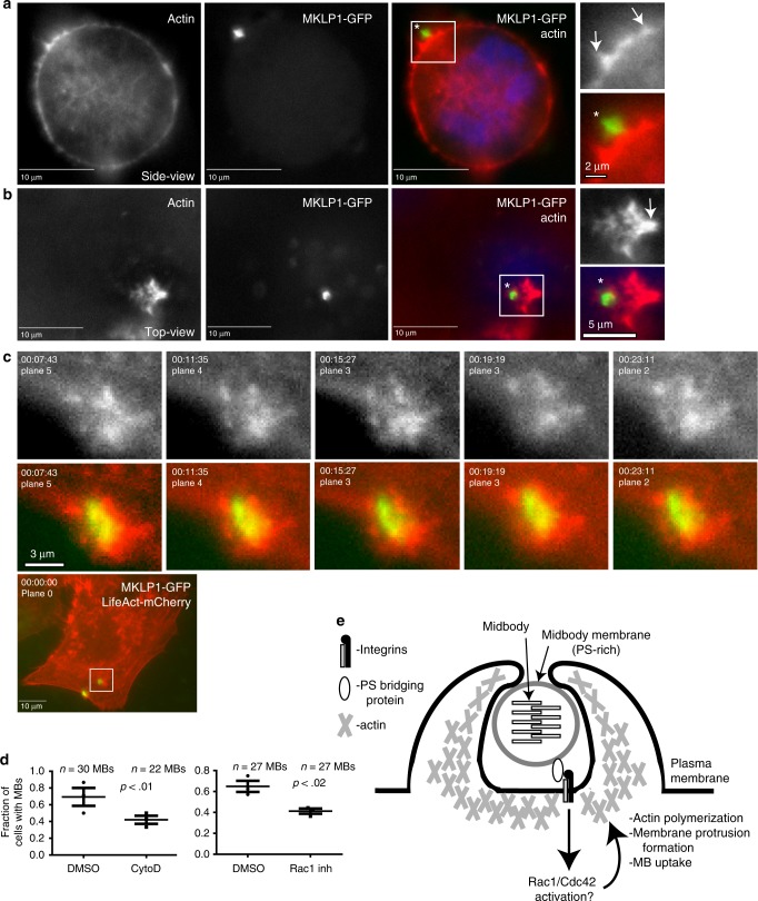
Actin polymerization mediates phagocytic MB internalization. a–c HeLa cells stably expressing LifeAct-mCherry were incubated with GFP-MBs for 3 h. Cells were then washed and analyzed by microscopy. Arrows in a and b point to actin-rich protrusions surrounding MBs during internalization. c Shows time-lapse series of actin rosette dynamics during MB internalization. d HeLa cells stably expressing LifeAct-mCherry were incubated with GFP-MBs for 3 h in the presence or absence of 10 μm of cytochalasin D or 100 μm of Rac1 inhibitor. Cells were then washed and incubated for another 24 h (without cytochalasin D or Rac1 inhibitor). The number of cells with internalized post-mitotic MBs were determined by fluorescence microscopy. The data shown are means and standard deviations derived from three independent experiments. n shows the number of cells analyzed for each experimental condition (Student’s unpaired, two-tailed t-test). e Schematic representation of proposed mechanism for MB internalization

