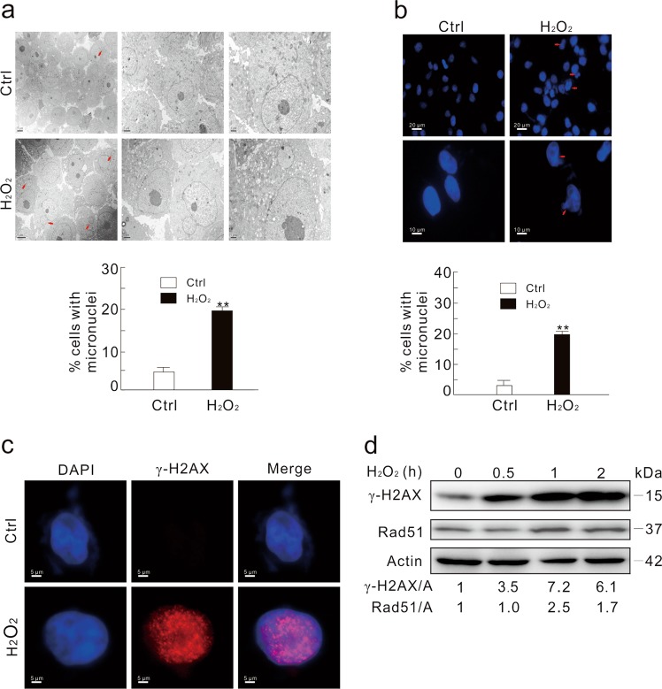
Fig. 1. H2O2 increases micronuclei formation.
a Electron microscopy was performed using the vehicle (Ctrl) or H2O2-treated (0.5 mM; 2 h) HeLa cells. The arrows indicate typical micronuclei. The number of cells containing micronuclei was counted and at least 30 cells were included in each group. b HeLa cells were treated with 0.5 mM H2O2 for 2 h, stained with DAPI, and observed with fluorescent microscope. The number of cells containing micronuclei was counted and at least 60 cells were included in each group. The data were normally distributed and statistically analyzed using the Studen–Newman–Keuls test. The double asterisks denote significant difference from control (**P < 0.01). c Immunofluorescence was performed using the γ-H2AX antibody in HeLa cells following treatment with 0.5 mM H2O2 for 2 h. d HeLa cells were treated with 0.5 mM H2O2 for the indicated times, and cell lysates were subjected to immunoblotting with the antibodies indicated. The adjusted ratios of γ-H2AX and Rad51 to actin (A) were presented below the blots. Similar experiments were repeated at least three times

