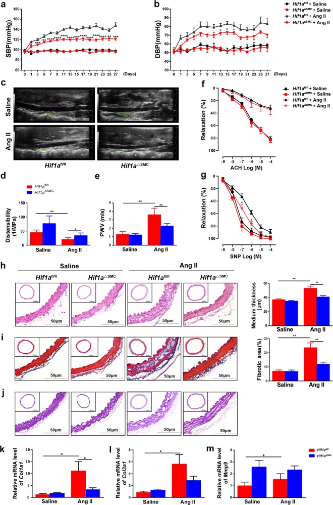
Fig. 2. HIF1α deficiency in SMCs suppresses Ang II-induced vascular remodeling in mice.
Hif1afl/fl and Hif1aΔSMC mice were infused with saline or Ang II (1000 ng/kg/min) for 28 days. a SBP and b DBP were measured by the tail-cuff method. *P < 0.05, **P < 0.01, ***P < 0.001 vs. Hif1afl/fl + Ang II; n = 10 per group, statistical significance was determined by two-way ANOVA analysis. M-mode ultrasound of abdominal aorta was acquired c, and the distensibility (d) as well as pulse wave velocity (PWV) (e) were measured. *P < 0.05, **P < 0.01, n = 6 per group. f, g Concentration–response curves of endothelium-dependent (acetylcholine, Ach) and endothelium-independent (sodium nitroprusside, SNP) relaxation. *P < 0.05 vs. Hif1afl/fl + Ang II, n = 6 per group. h Representative images of H&E staining and the mean medium thickness for the aortas. i Representative images of Masson’s trichrome staining and the fibrotic area of each group were analyzed. j Representative images of Elastin staining for the aortas. *P < 0.05, **P < 0.01, n = 8/saline group, n = 12/Ang II group. k–m Aortic Col1a1, Col3a1, and Mmp9 mRNAs were measured by qPCR. *P < 0.05, n = 6 per group. Statistical significance was determined by one-way ANOVA test followed by the unpaired t-test

