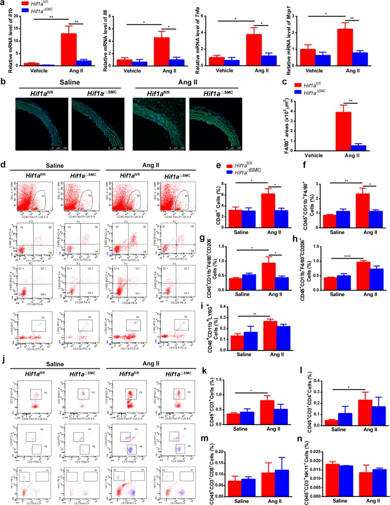
Fig. 3. SMC-specific HIF1α deficiency abolishes Ang II-induced M1 macrophage infiltration and vascular inflammation.
Hif1afl/fl and Hif1aΔSMC mice were infused with saline or 1000 ng/kg/min Ang II for 28 days. a Il1b, Il6, Tnfa, Mcp1 mRNAs in aortas after saline or angiotensin II infusion for 28 days were measured by qPCR. *P < 0.05, **P < 0.01, n = 6 per group. b Immunofluorescence staining of representative cross-sections of mice aortas for the F4/80-positive (green) cells and quantification (c) *P < 0.05, n = 6, statistical significance was determined by the unpaired t-test. Flow cytometry analysis was performed for the aortas (d, j) and CD45+ cells (e), CD45+CD11b+F4/80+ macrophages (f), CD45+CD11b+F4/80+CD206− M1 macrophages (g), CD45+CD11b+F4/80+CD206+ M2 macrophages (h), CD45+CD11b+LY6G+ neutrophils (i), CD45+CD3+ T cells (k), CD45+CD3+CD4+ T cells (l),CD45+CD3+CD8+ T cells (m), and CD45+CD3+NK11+ NKT cells (n) were quantified, respectively. *P < 0.05, **P < 0.01, ***P < 0.001, n = 6 per group. Statistical significance was determined by one-way ANOVA test followed by the unpaired t-test

