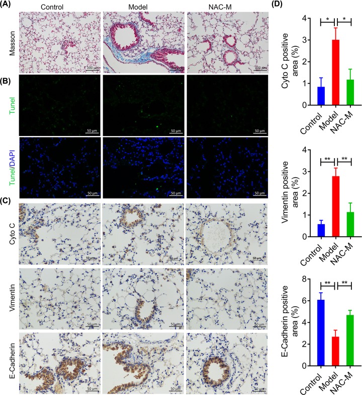
Figure 4. NAC administration alleviated the CS-induced tissue damages and pulmonary fibrosis in mice lungs.
(A) Representative images of Masson staining, measuring collagen deposition in lung tissues from CS-induced mice silicosis model following NAC administration for 5 months (20× magnification). (B) Representative images of TUNEL staining in the lungs tissues from CS-induced chronic pulmonary inflammation model mice following NAC administration for 5 months (40× magnification). (C-D) IHC analysis of the effect of NAC on the expression of Cyto C, E-cadherin and vimentin. *P<0.05, **P<0.01 versus model group.

