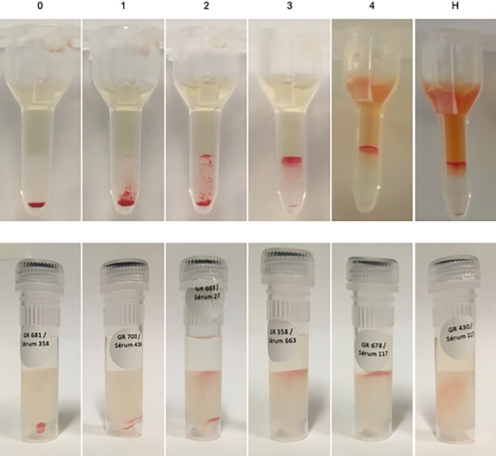
Figure 1.

Microgel and rapid gel grades. Agglutination grades for microgel assay (top panel) and rapid gel assay (bottom panel). 0: all erythrocytes passed through the gel and formed a compact pellet at the bottom, 1: most erythrocytes form a pellet at the bottom of the gel, but not compact, with few erythrocytes visible in the lower half of the gel, 2: erythrocytes are predominantly observed in the lower half of the gel column or are dispersed throughout the gel, 3: erythrocytes are dispersed on the top half of the gel with some retained on the gel surface, and 4: all erythrocytes are retained on top of the gel (here with some hemolysis in the microgel). Hemolysis (H) was considered present when red discoloration was observed in the microgel chamber (here with grade 4 agglutination) or in the gel itself for the rapid gel assay. In opposite to this rapid gel assay picture where only hemolysis can be appreciated, all 8 crossmatches in the current study with detectable hemolysis also resulted in positive agglutination (both with the rapid gel and microgel assays). Crossmatches with hemolysis or grades ≥2 agglutination were considered incompatible. For the top panel (microgel) grades 4 and H come from clinical cases of neonatal isoerythrolysis
