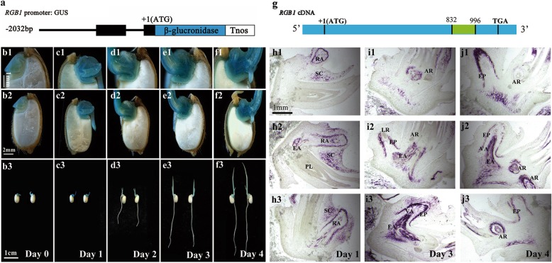
Fig. 2.
Expression pattern analysis of RGB1 in the embryo during early postgermination development in rice. (a) RGB1 promoter::GUS fusion gene. The 2.032-kb promoter region and the 105-bp fragment after ATG of RGB1 were used to drive the expression of the GUS gene. (b-f) Expression of the RGB1:GUS construct in median longitudinal sections of embryos. RGB1 was expressed in geminated seeds and young seedlings, strongly in the shoot-root axis. (b1-b3) Germinated seeds, (c1-c3) 1-day-old seedlings, (d1-d3) 2-day-old seedlings, (e1-e3) 3-day-old seedlings, (f1-f3) 4-day-old seedlings. (g) Location of the probe used for in situ hybridization of the RGB1 cDNA sequence (the fragment is shown in green). (h-j) In situ hybridization detection of RGB1 transcripts in rice embryos after germination. (h1-h3) Median longitudinal sections of 1-day-old wild-type seedlings. RA, radicle. SC, scutellum. EA, embryonic axis. PL, plumule. (i1-i3) Median longitudinal sections of 3-day-old wild-type seedlings. AR, adventitious root. EP, epidermis. EA, embryonic axis. VA, vascular tissue. LR, lateral root. (j1-j3) Median longitudinal sections of 4-day-old wild-type seedlings. AR, adventitious root. EP, epidermis. EA, embryonic axis. VA, vascular tissue. LR, lateral root

