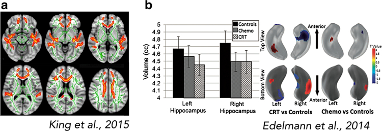
Fig. 6.

Altered brain structure in adult survivors of pediatric cancer. a Relative to matched controls (n = 27), adult survivors of childhood brain tumor (n = 27, ages 18–32; average of 13.7 years [SD = 5.37] since diagnosis) demonstrate reductions in indicators of white matter integrity in frontal and temporal areas. White matter integrity in frontal areas was positively correlated with IQ (King et al. 2015b). b Altered hippocampal volume and shape in adult survivors of pediatric ALL treated with CRT (n = 39, mean age = 26.7 years [SD = 3.4]; average of 23.9 years [SD = 3.1] since diagnosis) or CT only (n = 36, mean age = 24.9 years [SD = 3.6]; average of 15 years [SD = 1.7] since diagnosis) relative to controls (n = 23, mean age = 23.1 years; Edelmann et al. 2014). Abbreviations: ALL, acute lymphoblastic leukemia; CRT, cranial radiation therapy; CT, chemotherapy. All images are adapted with permission
