FIGURE 1.

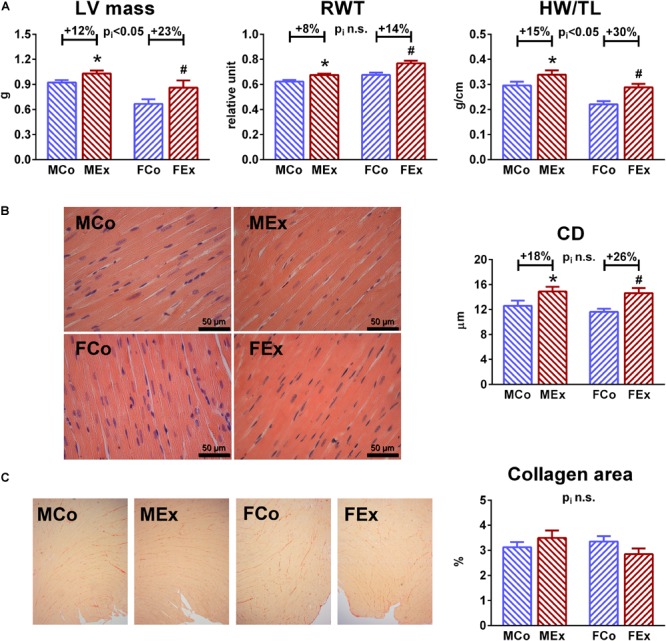
Characterization of exercise-induced left ventricular (LV) hypertrophy. (A) Echocardiographic LV mass and post-mortem measured heart weight (HW, normalized to TL) showed increased values in male exercised (MEx) and female-exercised (FEx) animals compared to male control (MCo), and female control (FCo) rats, respectively. Female gender was associated with greater degree of hypertrophy. A slight, but significant increase of relative wall thickness (RWT) suggest concentric type of hypertrophy in our rat model. (B) Representative hematoxylin-eosin stained sections (magnification 400 ×) from all of the groups, that were used to measure transnuclear cardiomyocyte width. Mean LV cardiomyocyte diameter (CD) was increased in both genders, that confirmed hypertrophy at cellular level. (C) One-one representative picrosirius-stained section from each group. Red color indicates collagen fibers, magnification 50 ×. Picrosirius-staining showed unaltered collagen density in exercised rats nor in male neither in female animals. Values are means ± SEM. ∗p < 0.05 vs. MCo, #p < 0.05 vs. FCo, pi: interaction p-value of two-way analysis of variance (ANOVA).
