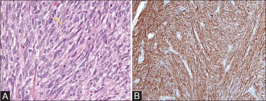
Figure 1 (A and B).

(A) H and E, ×40; Cellular proliferation of spindle cells with pale to eosinophilic fibrillar cytoplasm, arranged in whorls or short intersecting fascicles. Rare mitotic activity (arrow) (B) IHC, ×40; Immunohistochemistry for CD117 (CKIT) shows strong and diffuse cytoplasmic staining
