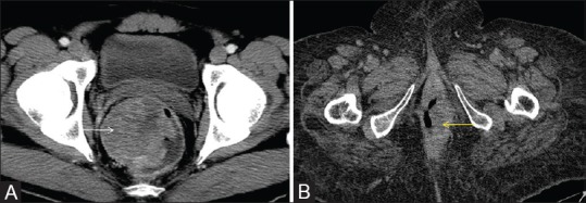
Figure 10 (A and B).

Axial post contrast CT showing (A) rectal GIST with intraluminal extension and showing heterogeneous post contrast enhancement (white arrow) and (B) anal canal GIST with intraluminal extension (yellow arrow)

Axial post contrast CT showing (A) rectal GIST with intraluminal extension and showing heterogeneous post contrast enhancement (white arrow) and (B) anal canal GIST with intraluminal extension (yellow arrow)