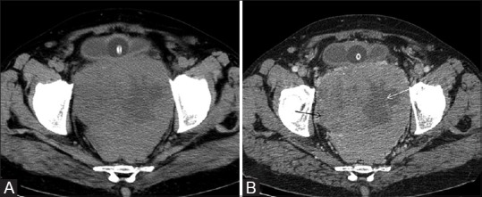
Figure 2 (A and B).

(A), pre contrast CT scan showing rectal GIST with heterogeneous attenuation with hypoattenuating areas within. (B) Post contrast CT scan heterogeneous GIST arising from the rectum with heterogeneous enhancement with non-enhancing cystic/necrotic areas within (arrows). It shows extraserosal extension anteriorly and abuts the urinary bladder without infiltration
