Figure 3.

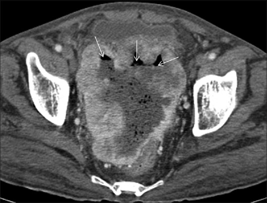
Post contrast axial CT scan showing sigmoid GIST with peripheral post contrast enhancement. Central non enhancing cystic/necrotic area is seen. Air fluid levels (arrows) are also noted within suggestive of communication with gut

Post contrast axial CT scan showing sigmoid GIST with peripheral post contrast enhancement. Central non enhancing cystic/necrotic area is seen. Air fluid levels (arrows) are also noted within suggestive of communication with gut