
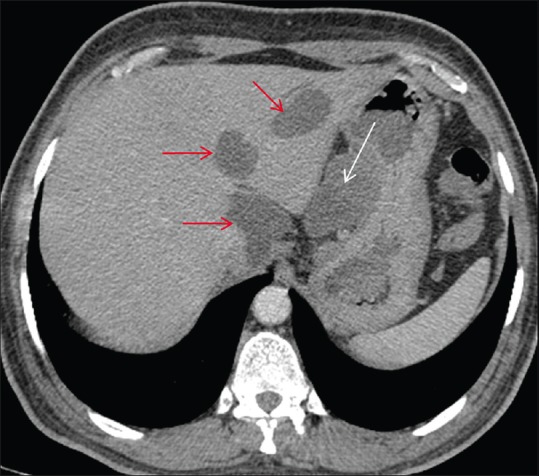
Figure 6.
Axial post contrast CT scan showing homogeneously enhancing exophytic gastric mass in the gastrohepatic region (white arrow) with few hypodense metastatic liver lesions (red arrow)
Official websites use .gov
A
.gov website belongs to an official
government organization in the United States.
Secure .gov websites use HTTPS
A lock (
) or https:// means you've safely
connected to the .gov website. Share sensitive
information only on official, secure websites.
Axial post contrast CT scan showing homogeneously enhancing exophytic gastric mass in the gastrohepatic region (white arrow) with few hypodense metastatic liver lesions (red arrow)